Investigation: An up-close look at the unique and complex appendages of an aquatic hermit crab
Crustaceans are complex invertebrates with highly specialised and unique body parts which have specific functions. Each segment has a pair of biramous appendages, many of which differ substantially in morphology compared to other body segments.
After watching a Blue striped hermit crab feed, a multitude of mouth parts could be seen rapidly moving, working together to deliver the food to the crab's mouth. This investigation is based on trying to decipher the physical characteristics and function of many of these appendages using a microscope and literature. Photographs were taken using either a camera or a Dinoscope microscope camera. The literature used to identify the body parts and their functions was Tudge, Asakura & Ahyong, (2004.) Two crabs were obtained for this study from the mudflats near Brighton Park on the Brisbane's North side, and dissected in the laboratory.
The first head appendage of a hermit crab is the antennulae, (see Figure 9,) a small pair of antenna which sit in between the two larger antenna. Two flagella can be seen at the distal end of this appendage, the largest of the two bears aesthetascs, which are chemosensory organs. Setae are present on the flagella, and the basal segment (not shown) bears a statocyst.
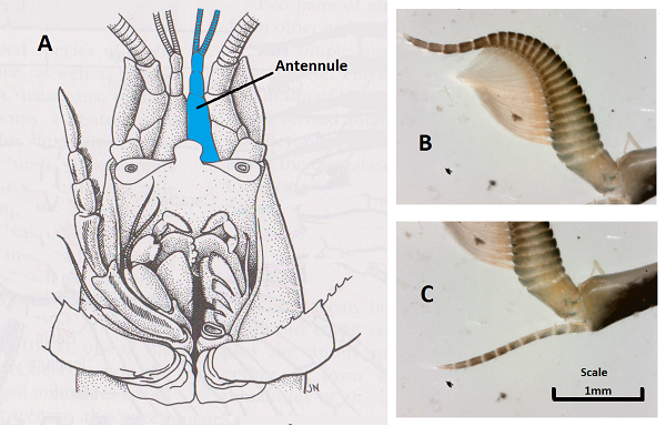
Figure 9- The Antennulae of Clibanarius longitarus. A: Typical crustacean head appendages (lobster) showing the location of the antennulae. B & C: Distal end of Clibanarius longitarus antennulae. (Source: Photographs by Author, 2014; diagram modified from Ruppert, Fox & Barnes, 2004)
Probably the most obvious head appendage of an aquatic hermit crab is the antenna, (see Figure 10,) bearing a single, long, uniramous flagellum. This happens to be the exopod of this appendage.
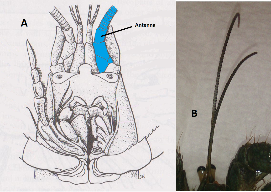
Figure 10- The antennae of Clibanarius longitarus. A: Typical crustacean head appendages (lobster) showing the location of the antennae. B: The antennae of Clibanarius longitarus. (Source: Photograph by Author, 2014; diagram modified from Ruppert, Fox & Barnes, 2004)
The third cephalic pair of appendages is the mandibles and their palps. Unfortunately, the palps could not be identified or photographed in this investigation. The mandibles themselves however were easy to find, (see Figure 11.) The incisor and molar processes are both calcarious, but the molar processes stand out with their slightly bigger size and white colouration. They also sit in front of the incisor processes, shielding them.
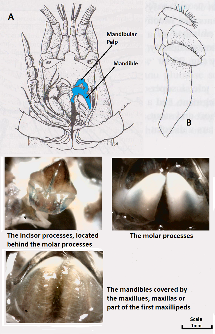
Figure 11- The mandibles of Clibanarius longitarus. A: Typical crustacean head appendages (lobster) showing the location of the mandibles. B: Typical paguroidea mandible and associated palp. (Source: Photographs by Author, 2014; diagrams modified from Ruppert, Fox & Barnes, 2004, and Tudge, Asakura & Ahyong, 2004)
The maxillules, maxilla, and first maxillipeds again, could not be identified. Some appendages were photographed but could not be identified with absolute confidence, so they will not be shown in this investigation. These appendages are the second, third and fourth mouthpart appendages, and would sit over, and slightly below the mandibles. All are flattened and bear setae, the first maxillipeds also bear a short flagellum. The maxilla function in creating a respiratory current, and the first maxillipeds aid in feeding.
The second maxillipeds bear a long flagellum on the exopod, and the endopods have well developed segments and are covered in setae, (see Figure 12.) The third maxillipeds are very similar, only slightly larger, (see Figure 13.) They are the largest of the mouth appendages in this species, and both play an important role in feeding.
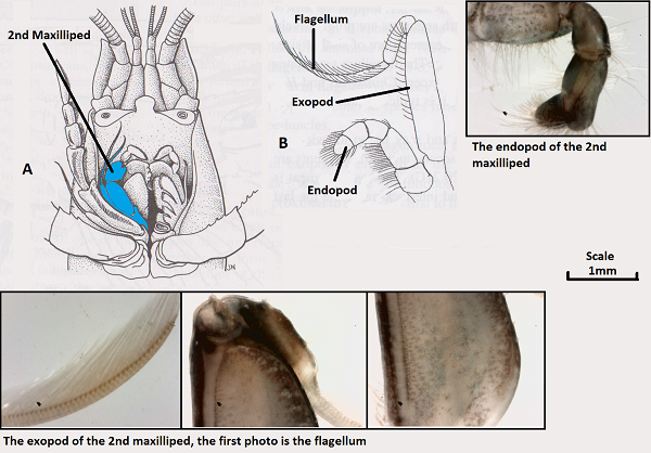
Figure 12- The second maxillipeds of Clibanarius longitarus. A: Typical crustacean head appendages (lobster) showing the location of the second maxillipeds. B: Typical paguroidea second maxillipeds showing the endopod, exopod, and flagellum. (Source: Photographs by Author, 2014; diagrams modified from Ruppert, Fox & Barnes, 2004, and Tudge, Asakura & Ahyong, 2004)
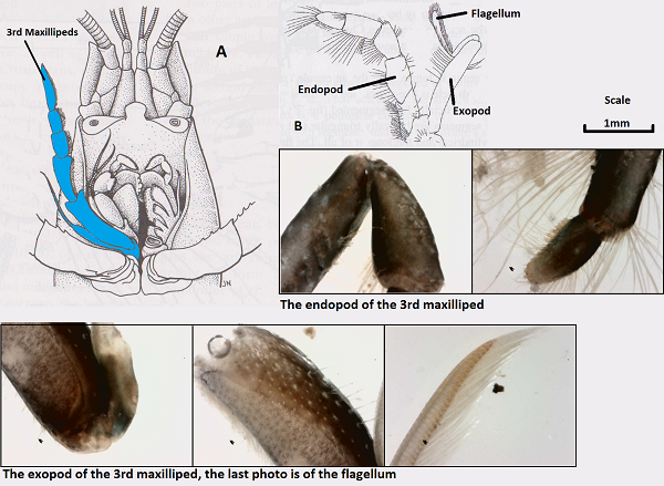
Figure 13- The third maxillipeds of Clibanarius longitarus. A: Typical crustacean head appendages (lobster) showing the location of the third maxillipeds. B: Typical paguroidea third maxillipeds showing the endopod, exopod, and flagellum. (Source: Photographs by Author, 2014; diagrams modified from Ruppert, Fox & Barnes, 2004, and Tudge, Asakura & Ahyong, 2004)
The fourth and fifth pereiopods are interesting appendages for hermit crabs because they are physically different to other crustacean species in that they are specialised to fit inside a shell. This means that they are reduced, and are heavily setosed to increase their grip on the inside of the shell. These pereiopods were photographed, and both of these morphological differences can clearly be seen, (see Figure 14.) A small hook-like structure can also be seen on the fourth pereiopod, which is not unusual for members of the paguroidea superfamily.
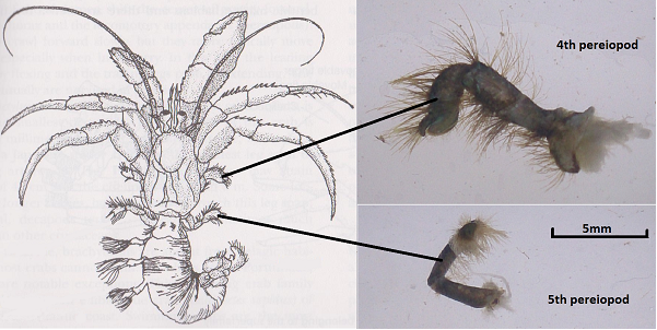
Figure 14- The smaller and heavily setosed fourth and fifth pereiopods of Clibanarius longitarus. (Source: Photographs by Author, 2014; diagrams modified from Ruppert, Fox & Barnes, 2004)
To further investigate the various body parts of this species, more photographs were taken and the best quality ones were published below, (see Figure 15.) They include eggs found in the pleon, a gill, the end of one of the walking legs or pereiopods, the dactyl of a cheliped, a compound eye and finally the uropods located on the telson. Another photo was taken without the microscope, of the crab's body with the carapace removed, revealing the gills, (see Figure 16.)
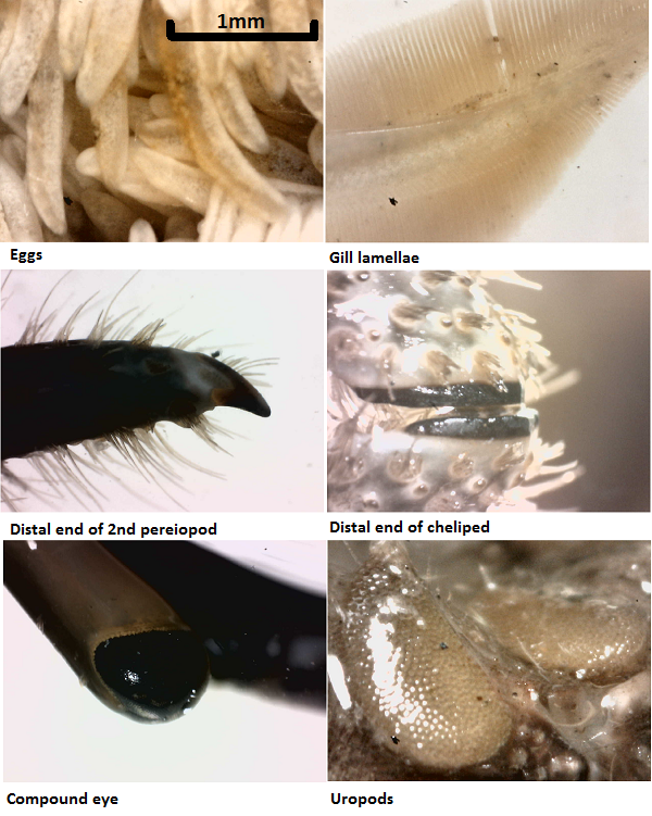
Figure 15- Other various body parts of Clibanarius longitarus. (Source: Photographs by Author, 2014)
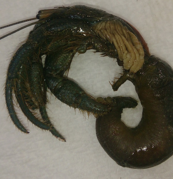
Figure 16- The exposed gills of Clibanarius longitarus as a result of the carapace being removed. (Source: Photograph by Author, 2014) |